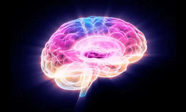
In a laboratory looking closely at the hippocampus, the part of the brain central to learning and memory, researchers have identified a striking shift tied to the aging brain. In mice, one protein kept standing out as the years passed: FTL1.
What did scientists find in the aging brain?
Scientists at UC San Francisco tracked changes in genes and proteins in the hippocampus of mice over time, looking for what set young animals apart from older ones. Among everything they examined, FTL1 was the only protein that appeared consistently different.
Older mice had higher levels of FTL1. At the same time, they showed fewer connections between neurons in the hippocampus and performed worse on cognitive tests. The pattern suggested more than simple wear and tear; it pointed to a specific biological change inside the aging brain.
When the team increased FTL1 in young mice, the effect moved quickly in the wrong direction. Their brains began to resemble those of older mice, and their behavior followed. In cell experiments, nerve cells engineered to produce high amounts of FTL1 developed simplified structures, forming short, single extensions instead of the complex branching networks seen in healthy cells.
Can lowering FTL1 improve the aging brain?
The most notable result came when researchers reduced FTL1 in older mice. Brain-cell connections increased, and memory test performance improved. Saul Villeda, PhD, associate director of the UCSF Bakar Aging Research Institute and senior author of the paper in Nature Aging, called it “a reversal of impairments, ” adding that it is “much more than merely delaying or preventing symptoms. ”
That finding matters because it shifts the story from observation to intervention. The research suggests that the aging brain may not only be mapped more precisely, but in some settings may also respond when the protein tied to decline is lowered.
How does FTL1 affect brain cell energy?
The study also found a metabolism link. In older mice, higher levels of FTL1 slowed cellular metabolism in the hippocampus. When researchers treated those cells with a compound that boosts metabolism, the negative effects were prevented. That detail gives the work a broader human dimension: memory decline is not only about structure, but also about how brain cells use energy to function.
The findings may help explain why the aging brain can lose resilience even before more visible changes appear. They also suggest a path for future treatments that target FTL1 and its effects, though the work remains in mice and does not establish a human therapy.
Why does this matter beyond the lab?
The research lands alongside a second line of evidence that broadens the picture. A study of 18, 701 participants from 34 countries found that physical and social exposomal factors were linked to multimodal brain age. The analysis showed that aggregated exposome models explained far more variance than individual exposures, and that physical exposome was associated more with structural brain aging while social exposome was more strongly tied to functional brain aging.
That work also found that exposome burden accounted for higher risk of accelerated aging, underscoring the role of physical, social, and political inequities. Together, the two studies point to a layered reality: the aging brain is shaped both by biology inside cells and by the environments around people.
What comes next for treatment and prevention?
Villeda said the findings could pave the way for treatments that target FTL1 and counter its effects in the brain. He described the moment as “a hopeful time to be working on the biology of aging. ” The researchers’ next challenge is clear: determine whether the same reversal seen in mice can translate beyond the lab.
For now, the image is both sobering and hopeful. In the hippocampus, a protein named FTL1 appears to help drive decline. But in the same place where memory falters, scientists also saw signs that recovery may be possible in the aging brain.